

My wonderfully talented 4-year-old male Labrador Retriever succumbed to Pythiosis in 2006. This emerging tropical disease is silently killing dogs around the U.S.
Most practicing vets have never seen this disease, so the information available on this site will give you more options in the diagnosis and treatment of your sick pet.
Pythiosis is an emerging tropical disease that was not seen in the U.S. 10-20 years ago when most practicing veterinarians attended vet school. Unless your vet has personally experienced this disease, they will probably NOT RECOGNIZE its symptoms and most DO NOT KNOW about the simple blood test to detect its antibodies or the immunotherapy treatment.
Early intervention is CRITICAL with this infection. PAVLAB in Texas has the fastest and easiest blood test available for both Pythiosis and Langenidium which is closely related. The PavLab immunotherapy treatment continues to be the least invasive and most effective treatment option. Contact them at 512-964-3927 for information.
PYTHIOSIS ALERTS (States where Pythiosis has been diagnosed)
Alabama, Arkansas, Arizona, California, Connecticut, Delaware, Florida, Georgia, Illinois, Kentucky, Louisiana, Maryland, Michigan, Missouri, Mississippi, New Jersey, North Carolina, Oklahoma, South Carolina, Tennessee, Texas, Wisconsin and Virginia. Bermuda.
Join our PYTHIOSIS group on Facebook to talk to others fighting this disease!
“Feel free to contact me anytime for additional information,” Tammy Bell
|
Dogs lost to Pythiosis
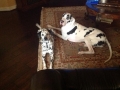                              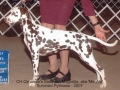                 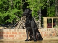           
Bella  Dude  Remi  Apollo 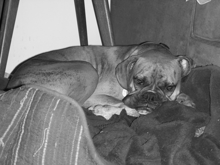 Beau 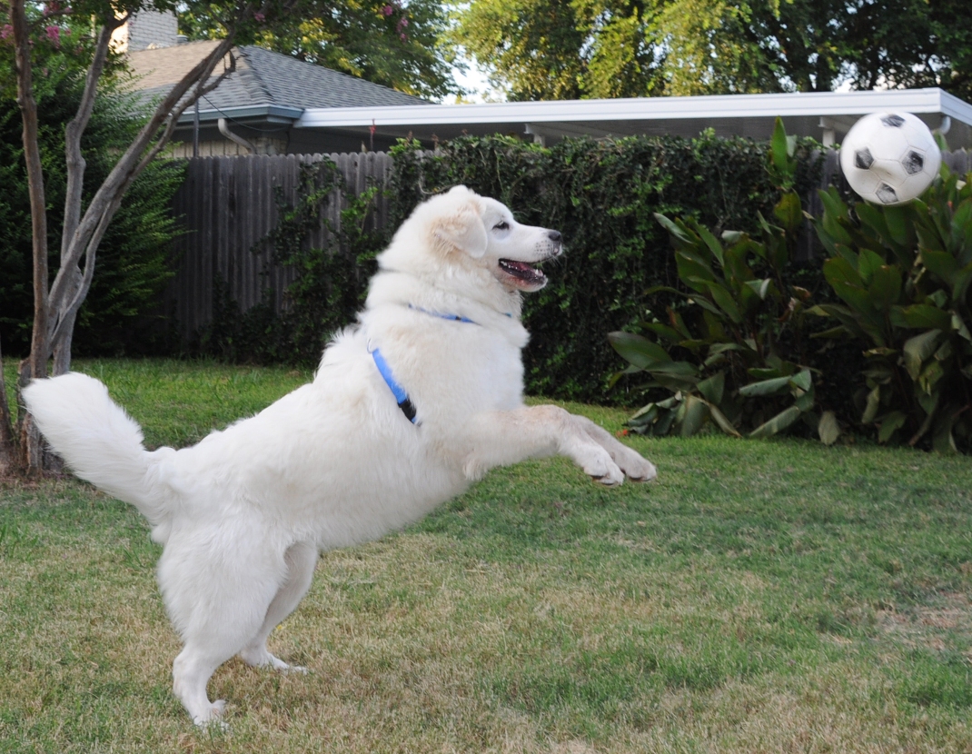 Bones 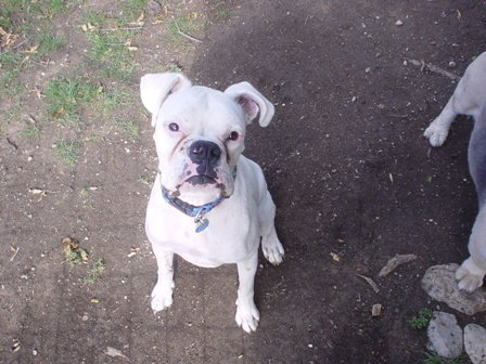 Bonnie 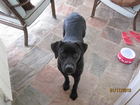 Callie  Charlie  Chestnut 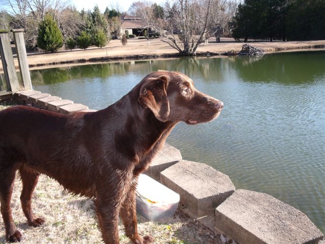 Chunk  Chunk 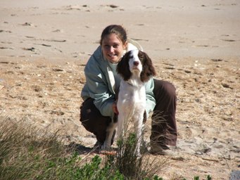 Coco 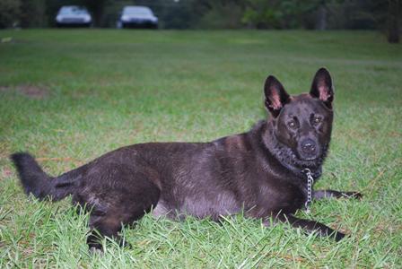 Gibson  Gretchen  Hannah   Hudson  Jive  Jordee  Koda  Lenny  Lu  Lu   Luke  Shep  Remy  Maggie  Posit 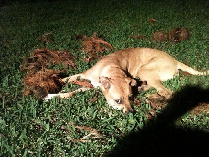 Maggie 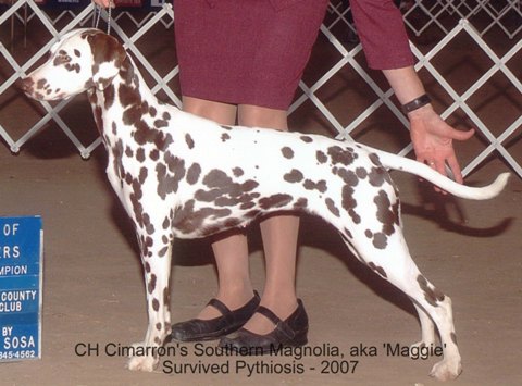 Marley  Molly  Montana  Murphy  Molly 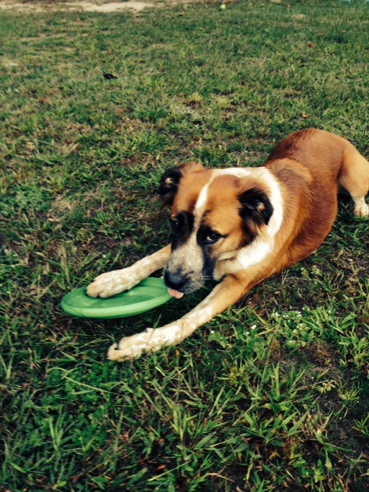 Leo  Osito   Duke  Rocky  Dory  Crimson  Ruckus 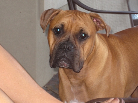 Ruger  Rusti 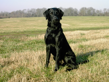 Rusti 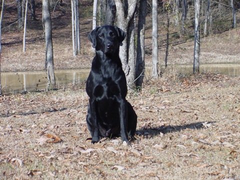 Rusti 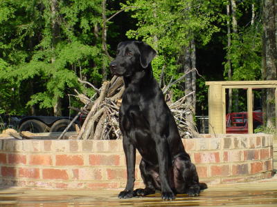 buster  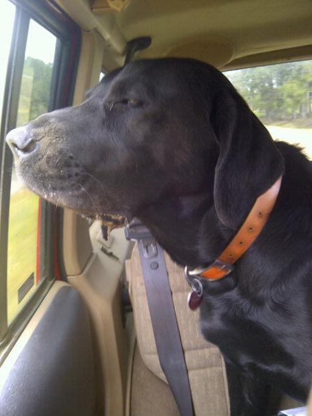      GE DIGITAL CAMERA   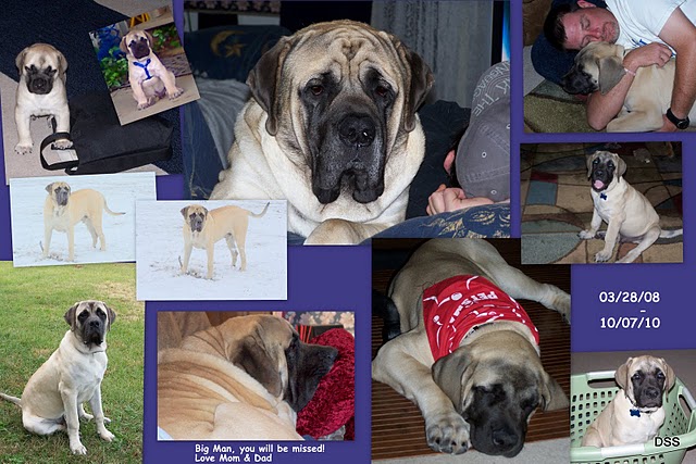  |

My father’s 2-3 year old black lab, Zeus, succumbed to pythiosis in 2013. He contracted it from a pond near the Texas coast. He was a healthy young dog that completely wasted away in just a couple short months. No lesions on his skin complicates the diagnosis. My mother who walked him and fed him everyday has been a veterinarian for 40 years and even she had trouble with the diagnosis until she contacted specialist colleagues.
By the time he was properly diagnosed and the treatments started it was too late. He was a wonderful dog and it’s mystifying we do not know more about the disease or it’s treatment.
Recently my black lab manifested gastrointestinal symptoms after swimming near the coast and I began frantically searching for info in pythiosis again. Fortunately it seems he just had a stomach bug and is rapidly improving.
I am glad to see your site and the fact that it comes up on the first page of google. Rusti was a beautiful dog and I’m extremely empathetic with what you had to go through. If you would like to add Zeus’ picture and/ or story to your site, I think that would be a good tribute to him. I’ve attached pictures below. Thank you for your time and effort.
Jeff Huebner
Austin, TX


Good evening Tammy,
I just came across your website. It made me feel a lot better knowing that my poor baby and I weren’t the only victims of this rare disease. My German Shepherd Ace has just turned 2; hadn’t even had the chance to truly experience life. He had this perfectly round lump on his left thigh and it grew almost half an inch in diameter every day or so…. Vets did a lot of unnecessary testing when, like you said, it’s just one simple blood test that will detect pythiosis. It had gotten to be the size of a cantaloupe when the vet broke the news to us that it had gotten too bad, even for amputation. He was put to rest this October……
I really appreciate your website and the group you’ve created for fellow victims. It’s really informative as it is quite hard to find information since pythiosis is so rare.
Madelyn
Columbia, South Carolina
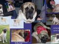
Here are some pictures of my darling Ace.

